Red and near-infrared light therapy shows remarkable healing effects across diverse populations, yet clinical outcomes vary dramatically between individuals. New research suggests melanin—long viewed merely as a passive optical filter—may actively modulate how therapeutic light penetrates and energizes cellular machinery.
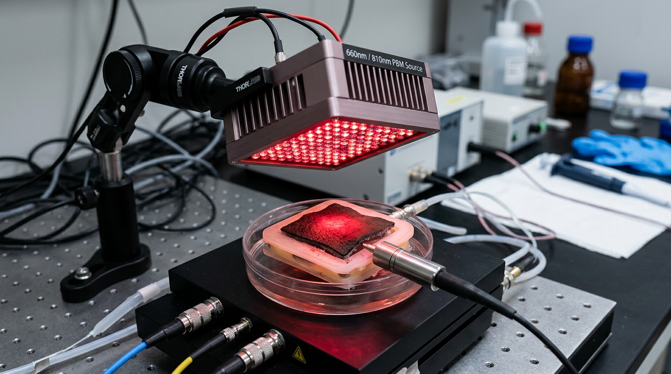
The phenomenon seems almost too simple to be profound: shine red light on injured tissue, and healing accelerates. Photobiomodulation (PBM) therapy, using wavelengths between 660-850 nanometers, has demonstrated efficacy in wound healing, pain reduction, and tissue regeneration across hundreds of clinical studies. Yet beneath this apparent simplicity lies a complex interplay between photons, cellular photoacceptors, and the often-overlooked optical properties of melanin.
Most PBM research has focused on cytochrome c oxidase (CCO), the terminal enzyme in the mitochondrial electron transport chain, as the primary photoacceptor. This copper-containing enzyme absorbs red and near-infrared light, leading to increased ATP production and downstream cellular signaling. However, this model—developed largely from studies on lightly pigmented tissues—may tell only part of the story.
The Melanin Absorption Paradox
Melanin's interaction with therapeutic light wavelengths presents a fascinating paradox. Eumelanin, the dominant form in human skin, exhibits broadband absorption that decreases monotonically from UV through the visible spectrum into the near-infrared. At 660 nm, a common therapeutic wavelength, melanin still absorbs significantly—approximately 10-fold higher than at 850 nm.
Traditional thinking suggested this absorption would simply attenuate therapeutic light, requiring higher doses for darker skin. However, recent investigations reveal a more nuanced picture. Research by Solís Herrera and colleagues has demonstrated that melanin can act as a photosensitizer, capable of dissociating water molecules when exposed to light and generating reducing equivalents that could complement mitochondrial energy production.
This finding challenges the binary view of melanin as either protective or problematic for light therapy. Instead, melanin-rich tissues may represent a distinct photobiological environment where multiple photoacceptor systems operate simultaneously. The hydrated melanin granules in melanosomes could serve as auxiliary energy conversion centers, potentially explaining why some individuals with higher melanin content show robust responses to PBM despite theoretical light attenuation.
Depth-Dependent Light Interactions
The spatial distribution of melanin within tissue creates complex optical environments that influence therapeutic light penetration. In human skin, melanin concentration varies dramatically with depth—highly concentrated in the basal epidermis (0.1-0.2 mm deep) but virtually absent in the deeper dermis and subcutaneous layers where many therapeutic targets reside.
Monte Carlo simulations of light transport in melanin-rich skin reveal that while surface absorption is indeed higher, the wavelength-dependent attenuation creates distinct therapeutic windows. At 810-850 nm, where melanin absorption is relatively low, therapeutic light can still reach depths of 2-4 cm in darker skin types, sufficient to influence dermal fibroblasts, muscle tissue, and even bone cells.
More intriguingly, the melanin absorption gradient may create beneficial photobiological effects at intermediate tissue depths. As therapeutic light encounters decreasing melanin concentrations with depth, the local photon flux increases, potentially creating zones of enhanced photobiomodulation activity. This could explain clinical observations of robust wound healing responses in individuals with higher melanin content, despite theoretical predictions of reduced light penetration.
Cellular Signaling Beyond Mitochondria
Recent research has expanded our understanding of PBM mechanisms beyond the mitochondrial paradigm. Ion channels, particularly calcium and potassium channels, show light sensitivity in the red and near-infrared spectrum. The bioelectric properties of cells—their membrane potential and ion flux patterns—can be directly influenced by therapeutic light exposure.
Melanin's role in this expanded model becomes particularly interesting when considering its semiconductor properties. With a bandgap of approximately 1.85 eV, melanin can be photoexcited by photons in the therapeutic wavelength range. This photoexcitation could influence local electric fields and ion channel behavior, creating a secondary pathway for light-induced cellular responses.
Furthermore, melanin's stable free radical content, detectable by electron paramagnetic resonance (EPR) spectroscopy, may interact with the reactive oxygen species (ROS) signaling pathways activated by PBM. Rather than simply scavenging ROS, melanin might modulate redox signaling in ways that enhance therapeutic outcomes—a hypothesis supported by observations of improved antioxidant responses in melanin-rich tissues following light therapy.
Clinical Implications and Personalized Dosimetry
Understanding melanin's active role in photobiomodulation has immediate clinical implications. Current dosimetry protocols often apply simple correction factors based on skin phototype, essentially treating melanin as a neutral density filter. However, if melanin actively participates in therapeutic light responses, optimal dosing strategies may be more complex.
Clinical studies comparing PBM outcomes across different skin types have yielded mixed results, with some showing reduced efficacy in darker skin and others showing equivalent or even enhanced responses. These apparently contradictory findings may reflect the dual nature of melanin's interaction with therapeutic light—both attenuating and potentially amplifying photobiological responses through distinct mechanisms.
The emerging picture suggests that personalized photobiomodulation protocols should account not just for melanin content but for its distribution, hydration state, and local tissue environment. Advanced dosimetry might incorporate real-time feedback from bioelectric measurements or spectroscopic assessment of melanin photoexcitation to optimize therapeutic outcomes.
Key Takeaways
• Melanin acts as more than a passive optical filter in photobiomodulation therapy, potentially serving as an auxiliary photoacceptor system that complements cytochrome c oxidase-mediated responses.
• The wavelength-dependent absorption properties of melanin create complex therapeutic windows, with 810-850 nm light showing optimal penetration while still maintaining biological activity in melanin-rich tissues.
• Melanin's semiconductor properties and stable free radical content may contribute to photobiomodulation effects through bioelectric modulation and redox signaling pathways independent of mitochondrial mechanisms.
• Clinical outcomes in photobiomodulation therapy may depend on the interplay between melanin distribution, tissue depth, and wavelength-specific absorption rather than simple light attenuation models.
• Personalized dosimetry protocols that account for melanin's active photobiological role could significantly improve therapeutic outcomes across diverse populations.
• The spatial gradient of melanin concentration in tissue may create zones of enhanced photobiomodulation activity at intermediate depths, explaining robust clinical responses in individuals with higher melanin content.
References
Hamblin, M. R. "Mechanisms and applications of the anti-inflammatory effects of photobiomodulation." AIMS Biophysics 4(3), 337-361 (2017). DOI: 10.3934/biophy.2017.3.337
Karu, T. "Primary and secondary mechanisms of action of visible to near-IR radiation on cells." Journal of Photochemistry and Photobiology B 49(1), 1-17 (1999). DOI: 10.1016/S1011-1344(98)00219-X
Solís-Herrera, A., et al. "The unexpected capacity of melanin to dissociate the water molecule fills the gap between the life before and after ATP." Biomedical Research 21(2), 224-226 (2010).
Zhai, H., et al. "Skin anti-inflammatory agents: An overview." Skin Pharmacology and Physiology 17(4), 143-152 (2004). DOI: 10.1159/000078824
Chung, H., et al. "The nuts and bolts of low-level laser (light) therapy." Annals of Biomedical Engineering 40(2), 516-533 (2012). DOI: 10.1007/s10439-011-0454-7
Mester, E., et al. "Effect of laser rays on wound healing." The American Journal of Surgery 122(4), 532-535 (1971). DOI: 10.1016/0002-9610(71)90482-X