Recent research reveals that melanin, long studied primarily for its photoprotective role, may fundamentally influence the bioelectric patterns that govern cellular behavior. This emerging connection between pigment biochemistry and membrane voltage regulation suggests melanin operates as more than a passive shield — it may actively participate in the bioelectric signaling networks that control proliferation, differentiation, and tissue organization.
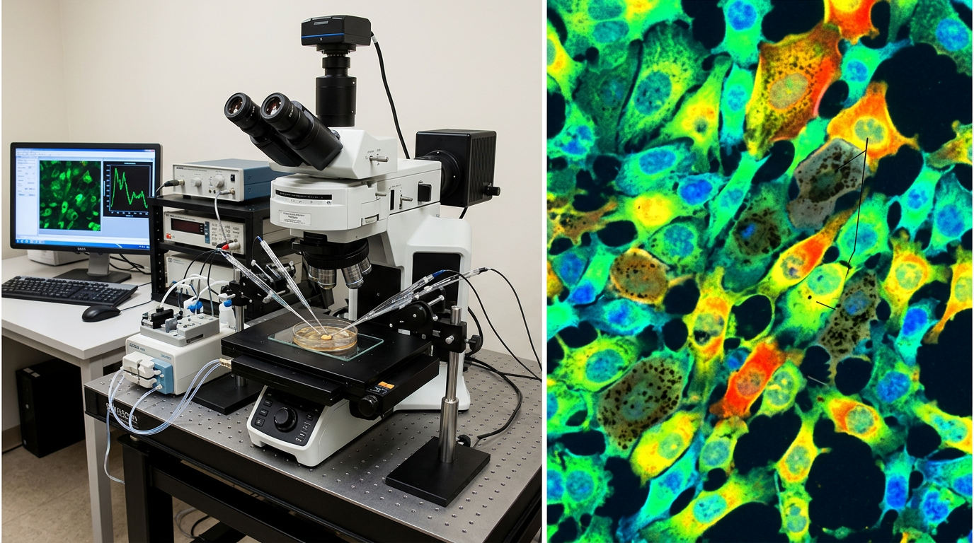
When developmental biologist Michael Levin's team at Tufts University demonstrated that cells maintaining membrane voltages below -20mV consistently exhibit proliferative, stem-like characteristics — and that this bioelectric signature appears in both embryonic development and cancer progression — they opened a new window into how life orchestrates its most fundamental processes. What they likely didn't anticipate was how this bioelectric framework might intersect with melanin biology in ways that could reshape our understanding of both fields.
The bioelectric code that Levin and colleagues have mapped operates through precise voltage gradients across cell membranes. Normal differentiated cells typically maintain membrane potentials around -50 to -70mV, while stem cells and cancer cells cluster in the depolarized range above -20mV. This isn't merely correlation — experimental manipulation of membrane voltage can drive cells toward proliferation or differentiation, suggesting that bioelectric patterns serve as a fundamental control mechanism for cellular identity.
Melanin's Electroactive Properties
Melanin's electrical characteristics have intrigued researchers since John McGinness first described its semiconductor behavior in 1974. Eumelanin, the brown-black pigment predominant in human skin and hair, exhibits a bandgap of approximately 1.85eV — positioning it as a wide-bandgap organic semiconductor. More remarkably, melanin's conductivity increases dramatically with hydration, suggesting it operates as a protonic conductor under physiological conditions.
This protonic conductivity mechanism distinguishes melanin from conventional electronic semiconductors. In hydrated states, melanin can facilitate proton movement along hydrogen-bonded networks within its polymer structure. Arturo Solís Herrera's research group has documented how melanin can split water molecules when exposed to electromagnetic radiation, generating proton gradients that could directly influence local pH and, consequently, membrane voltage dynamics.
The stable free radical character of melanin adds another layer of electroactive complexity. Electron paramagnetic resonance (EPR) spectroscopy consistently detects unpaired electrons in melanin samples — approximately 10^17 to 10^18 spins per gram in eumelanin. These stable radicals can participate in redox reactions and electron transfer processes that extend far beyond the melanin granule itself.
Membrane Voltage Modulation Mechanisms
The intersection of melanin's electroactive properties with cellular bioelectricity likely operates through multiple pathways. Ion channel modulation represents the most direct mechanism. Melanin's ability to chelate metal ions — particularly calcium, iron, and zinc — could influence the activity of voltage-gated channels that depend on these cofactors. Changes in local ion concentrations around melanin-containing organelles could shift the equilibrium potentials that determine membrane voltage.
Redox coupling offers another pathway for melanin-bioelectric interaction. The stable radicals in melanin can engage in electron transfer with cellular redox systems, potentially influencing the NADH/NAD+ ratios that drive many bioelectric processes. Research by Felix Scholkmann and others has shown that melanin can interact with mitochondrial electron transport chains, suggesting direct coupling between melanin redox states and cellular energetics.
The proton gradient hypothesis provides perhaps the most intriguing mechanism. If melanin generates localized proton gradients through its water-splitting activity, these pH changes could directly modulate membrane voltage through proton-coupled transport systems. Many ion channels and transporters exhibit pH sensitivity in the physiological range, making melanin's proton-generating capacity a potential voltage regulator.
Implications for Disease and Development
The melanin-bioelectric axis may illuminate puzzling aspects of melanoma biology. Melanoma cells often exhibit altered bioelectric properties compared to normal melanocytes, including changes in membrane potential and ion channel expression. If melanin normally contributes to maintaining appropriate membrane voltage in melanocytes, disruption of this system could contribute to the bioelectric signatures associated with malignant transformation.
This framework also suggests new perspectives on vitiligo and other pigmentary disorders. Beyond the obvious cosmetic effects of melanin loss, these conditions might involve disruption of bioelectric signaling in affected tissues. The observation that vitiligo patches sometimes exhibit altered wound healing and immune responses could reflect underlying bioelectric dysfunction rather than purely pigmentary changes.
In neuromelanin contexts, the bioelectric connection becomes even more compelling. The substantia nigra neurons that accumulate neuromelanin throughout life are precisely the cells that degenerate in Parkinson's disease. While iron-catalyzed oxidative stress has long been implicated in this neurodegeneration, the bioelectric properties of neuromelanin-containing neurons remain largely unexplored. Given that neuronal membrane potential directly controls neurotransmitter release and synaptic plasticity, melanin-mediated voltage modulation could influence dopaminergic function in ways we're only beginning to appreciate.
Future Research Directions
The melanin-bioelectric axis opens multiple avenues for investigation. Patch-clamp studies comparing membrane voltage dynamics in melanin-rich versus melanin-deficient cells could directly test whether pigment content influences bioelectric behavior. Such experiments would need to control for cell type and differentiation state while systematically varying melanin content.
Optogenetic approaches might allow researchers to manipulate melanin's photoactive properties while simultaneously monitoring membrane voltage changes. Since melanin absorbs across the visible spectrum, specific wavelengths could potentially drive different electroactive responses, providing a tool for dissecting mechanism.
The therapeutic implications deserve serious attention. If melanin does modulate membrane voltage, then bioelectric therapies targeting voltage-gated channels might need to account for pigmentation differences between patients. Conversely, melanin-based interventions might offer new approaches for bioelectric medicine, particularly in contexts where precise voltage control is therapeutically relevant.
Key Takeaways
• Melanin's semiconductor properties and protonic conductivity position it to influence cellular membrane voltage through multiple mechanisms including ion channel modulation and redox coupling.
• The -20mV depolarization threshold identified in Levin's bioelectric framework may be modulated by melanin content, potentially explaining some aspects of pigment cell biology and melanoma progression.
• Neuromelanin accumulation in dopaminergic neurons suggests a possible bioelectric component to Parkinson's disease pathogenesis beyond traditional oxidative stress models.
• Pigmentary disorders like vitiligo might involve bioelectric dysfunction in addition to cosmetic changes, opening new therapeutic approaches.
• The melanin-bioelectric axis represents a convergence of materials science, cell biology, and bioelectric medicine that could reshape our understanding of how pigment systems integrate with cellular signaling networks.
• Future research combining patch-clamp electrophysiology with melanin manipulation could directly test whether pigment content influences the bioelectric patterns that govern cellular behavior.
References
Levin, M. "Bioelectric signaling: Reprogrammable circuits underlying embryogenesis, regeneration, and cancer." Cell 184(8), 1971-1989 (2021). DOI: 10.1016/j.cell.2021.02.034
McGinness, J., Corry, P., & Proctor, P. "Amorphous semiconductor switching in melanins." Science 183(4127), 853-855 (1974). DOI: 10.1126/science.183.4127.853
Solís-Herrera, A., Arias-Esparza, M.C., & Solís-Arias, R.I. "The unexpected capacity of melanin to dissociate the water molecule fills the gap between the life before and after ATP." Biomedical Research 21(2), 224-226 (2010).
Mostert, A.B. "Melanin, the what, the why and the how: An introductory review for materials scientists interested in flexible and versatile polymers." Polymers 13(10), 1670 (2021). DOI: 10.3390/polym13101670
Blackiston, D.J., McLaughlin, K.A., & Levin, M. "Bioelectric controls of cell proliferation: Ion channels, membrane voltage and the cell cycle." Cell Cycle 8(21), 3527-3536 (2009). DOI: 10.4161/cc.8.21.9888
Zecca, L., Youdim, M.B., Riederer, P., Connor, J.R., & Crichton, R.R. "Iron, brain ageing and neurodegenerative disorders." Nature Reviews Neuroscience 5(11), 863-873 (2004). DOI: 10.1038/nrn1537